Mouse Interleukin-4 Recombinant
Categories: HematopoietinsRecombinant Mouse Cytokines$70.00 – $3,500.00
Description
Accession
P07750
Source
Optimized DNA sequence encoding Mouse Interleukin-4 mature chain was expressed in Escherichia Coli.
Molecular weight
Native Mouse Interleukin-4, generated by the proteolytic removal of the signal peptide and propeptide,the molecule has a calculated molecular mass of approximately 14 kDa. Recombinant Interleukin-4 is a monomer protein consisting of 121 amino acid residue subunits, migrates as an approximately 14 kDa protein under reducing conditions in SDS-PAGE.
Purity
>96%, as determined by SDS-PAGE and HPLC
Biological Activity
The ED(50) was determined by the dose-dependent stimulation of the proliferation of human TF-1 cells is ≤.4 ng/ml, corresponding to a specific activity of ≥x units/mg.
Protein Sequence
MGLNPQLVVI LLFFLECTRS HIHGCDKNHL REIIGILNEV TGEGTPCTEM DVPNVLTATK NTTESELVCR ASKVLRIFYL KHGKTPCLKK NSSVLMELQR LFRAFRCLDS SISCTMNESK STSLKDFLES LKSIMQMDYS
Endotoxin
Endotoxin content was assayed using a LAL gel clot method. Endotoxin level was found to be less than 0.1 ng/µg(1EU/µg).
Presentation
Interleukin-4 was lyophilized from a 0.2 μm filtered solution in PBS, pH.
Reconstitution
A quick spin of the vial followed by reconstitution in distilled water to a concentration not less than 0.1 mg/mL. This solution can then be diluted into other buffers.
Storage
The lyophilized protein is stable for at least years from date of receipt at -20° C. Upon reconstitution, this cytokine can be stored in working aliquots at2° -8° C for one month, or at -20° C for six months, with a carrier protein without detectable loss of activity. Avoid repeated freeze/thaw cycles.
Usage
This cytokine product is for research purposes only.It may not be used for therapeutics or diagnostic purposes.
Biological Process
Molecular function
Molecular function
Methods
In-vitro NK-DC co-culture
- Whole blood samples were obtained from healthy laboratory donors for the isolation of purified and enriched NK and DC cell populations from PBMC using magnetic cell separation.
- The development of an NK-DC co-culture system required two independent steps.
- Firstly, DC were prepared using a pre-optimised magnetic cell sorting kit for the extraction of CD14+ monocytes from PBMC.
- Following isolation, CD14+ monocytes were treated with 100 ng/ml of GM-CSF and 1000 U/ml of IL-4 in RPMI containing 5% autologous serum and maintained in a 5% CO2 cell culture incubator at 37°C for 5 days.
- This altered their phenotype to that of immature dendritic cells (iDC) with a population of >90% CD14−CD1a+ iDC cells, comparable with the published literature +CD16+ NK cell.
- Purities of >98% were attained consistent with published literature 2 cell culture incubator at 37°C.
- Co-culture cell ratios…
Primary cell isolation and viral infection
- Bone marrow macrophages (BMM) and bone marrow dendritic cells (BMDC) were generated as described previously
Immunoprotection analysis of Com1- and HspB-pulsed BMDCs
- Mouse bone marrow dendritic cells (BMDCs, CD11c+) were isolated from the bone marrow of BALB/c mice, according to the protocol described previously [2.
- Approximately 1 × 10⁶ cells was added to each well of a six-well plate, and mouse GM-CSF (20 ng/ml) and IL-4 (10 ng/ml) was added to the culture medium every other day.
- After 6 days of culture, BMDCs were stimulated with
C. - burnetii I Ag (10 μg/ml), Com1 (10 μg/ml), HspB (10 μg/ml),
E. - coli LPS (2 μg/ml), or 25 μl elution buffer (mock pulse) for 24 h at 37°C and 5% CO2.
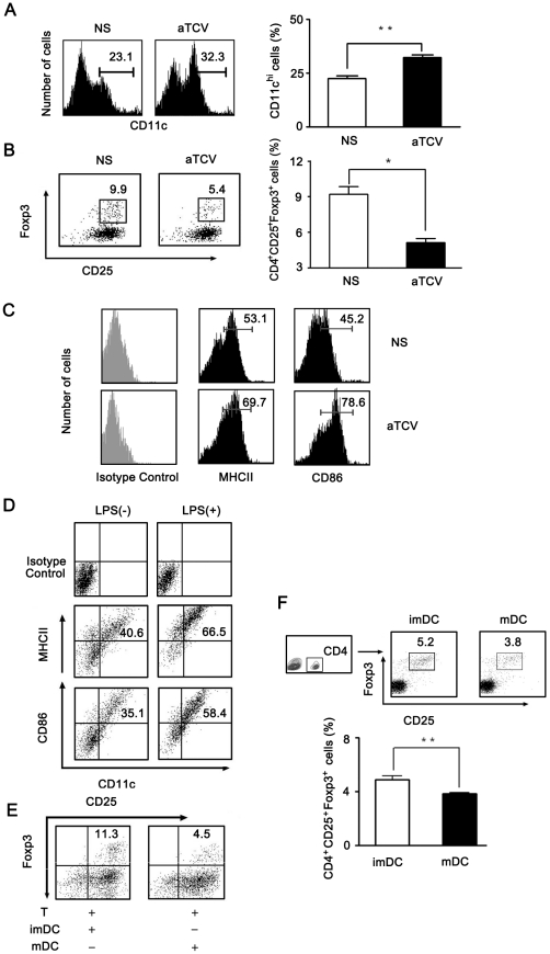
mDCs inhibited Treg differentiation.
- D–F, Bone marrow precursor cells freshly isolated from naïve mice were cultured in vitro in the presence of GM-CSF and IL-4.
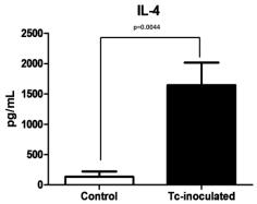
T. crassiceps-inoculation induces elevation of IL-4 and hepatic expression of Ym-1.
- Six weeks post-inoculation, IL-4 and IFN-γ serum levels were determined from Tc-inoculated and non-inoculated mice by ELISA.
Antibodies and proteins
- Antibodies directed against IκBα (#4814), phospho p38 (#9211), p38 (#9212), phospho ERK1/2 (#9106), ERK1/2 (#9102), phospho JNK1/2 (#9251), JNK1/2 (#9252) and phospho c-Jun (#3270) were all from Cell Signaling Technology.
- Wnt5a antibody was from Biotechnology (Sc-30224), NFATC2 for Western blotting was from and for immunofluorescence from (Sc-13034), and the mouse monoclonal β-actin from .
- LPS (from
E. - Recombinant IL-4 and IL-13 proteins were bought from .
- HlyA was obtained from Prof. S. Bhakdi (Mainz University, Germany).
- HlyA was preincubated with polymyxin B (50 µg/ml) at 4°C for 30 min to remove any possible LPS contamination.
Functional assays
- For cytokine signalling, LN T cells (5 × 10⁶ cells/ml) were cultured 16 h with recombinant mouse IL-7 (10 ng/ml), IL-4 (40 ng/ml), IL-6 (45 ng/ml& ) or IL-15 (100 ng/ml& ) as previously described (
Immune modulation by IL-12 shRNA silenced DC.
- Co-cultured T cells were harvested and gene expression of IFNγ, IL-2, IL-4 and IL-10 were detected by RT-PCR.
Spleen DC isolation and peripheral blood derived cultures
- After experimentation the following protocol, adapted from mammalian methods 2 flasks with phenolic caps at a density of approximately 5×10⁶ cells/ml of supplemented medium'>L-15 medium (20% FBS, 50 µg/mL gentamicin, and 0.25 µg/mL fungizone).
- Cells were allowed to adhere for two hours, at which point non-adherent cells were washed off and new media was added containing mammalian IL-4 (250 U/ml, human:, ; :, , ) and GM-CSF (1000 U/ml, human:,; :).
- Cells were “fed” additional IL-4 and GM-CSF (same concentration as above) on days two, four, and six.
- Non-adherent cells removed at the 2 hr time point were transferred to a new flask for culture without addition of exogenous IL-4 or GM-CSF (or removal of non-adherent cells).
- These mixed cultures (sans cytokines) yielded much the same result as the adherent culture with cytokines.
- A more direct comparison of adherent cultures with or without exogenous cytokines was made.
- No discernable…
Progressive acquisition of CD200R correlates with effector cytokine secretion.
- Intracellular cytokine staining with CD200R expression is shown for TNFα, IL-2, IL-4 and IFNγ.