Mouse Interleukin-6 Recombinant
Categories: HematopoietinsIL-6 gp130 familyRecombinant Mouse Cytokines$70.00 – $4,700.00
Description
Accession
P08505
Source
Optimized DNA sequence encoding Mouse Interleukin-6 mature chain was expressed in Escherichia Coli.
Molecular weight
Native Mouse Interleukin-6, generated by the proteolytic removal of the signal peptide and propeptide, the molecule has a calculated molecular mass of approximately 22kDa. Recombinant IL-6 is a monomer protein consisting of 122 amino acid residue subunits, and migrates as an approximately 22kDa protein under non-reducing and reducing conditions in SDS-PAGE.
Purity
>97%, as determined by SDS-PAGE and HPLC
Biological Activity
The ED(50) determined by the dose-dependent proliferation of murineTD1 cells, was found to be <.01 ng/ml cells was found to be in the range ofx108U/mg.
Protein Sequence
MKFLSARDFH PVAFLGLMLV TTTAFPTSQV RRGDFTEDTT PNRPVYTTSQ VGGLITHVLW EIVEMRKELC NGNSDCMNND DALAENNLKL PEIQRNDGCY QTGYNQEICL LKISSGLLEY HSYLEYMKNN LKDNKKDKAR VLQRDTETLI HIFNQEVKDL HKIVLPTPIS NALLTDKLES QKEWLRTKTI QFILKSLEEF LKVTLRSTRQ T
Endotoxin
Endotoxin content was assayed using a LAL gel clot method. Endotoxin level was found to be less than 0.1 ng/µg(1EU/µg).
Presentation
Recombinant Interleukin-6 was lyophilized from a 0.2 μm filtered PBS solution.
Reconstitution
A quick spin of the vial followed by reconstitution in distilled water to a concentration not less than 0.1 mg/mL. This solution can then be diluted into other buffers.
Storage
The lyophilized protein is stable for at least years from date of receipt at -20° C. Upon reconstitution, this cytokine can be stored in working aliquots at2° -8° C for one month, or at -20° C for six months, with a carrier protein without detectable loss of activity. Avoid repeated freeze/thaw cycles.
Usage
This cytokine product is for research purposes only.It may not be used for therapeutics or diagnostic purposes.
Biological Process
Molecular function
Molecular function
Methods
Materials
- Tetramethylammonium hydroxide was from Shanghai Lingfeng Chemical Reagent Co Ltd, Shanghai, China.
- Other materials were RPMI 1640 medium, penicillin, and streptomycin , fetal bovine serum (, , , the ), recombinant murine granulocyte-macrophage colony-stimulating factor, recombinant murine interleukin-4, CD11c, B7-1 (CD80), B7-2(CD86), MHC II, and chemokine receptor 7 (eBioscience, San Diego, CA), tumor necrosis factor-α, interleukin-1β, and interleukin-6 , prostaglandin E2 , a Prussian blue staining kit .
- Annexin V and propidium iodide , and the Cell Counting Kit-8 .
MAVS deficiency increases inflammatory cytokines in the lung of γHV68-infected mice.
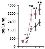
- Saline buffer, recombinant mouse IL6 or TNFα (rmIL6 or rmTNFα, 30 ng/mouse/day) was intranasally administered from 1 to 5 days post-infection (d.p.i.).
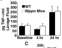
IL-6 is elevated in Nippo mice.
- Levels of TNF-α, IL-6, and IL-1β were measured by ELISA in peritoneal lavage fluid from WT or Nippo mice at baseline, 4 and 24 h after i.p.
Experimental agents
- Tetramethylammonium hydroxide was obtained from Lingfeng Chemical Reagent Co Ltd .
- medium'>RPMI medium 1640, penicillin and streptomycin were from .
- Fetal bovine serum (, , , the ), recombinant murine granulocyte-macrophage colony-stimulating factor, recombinant murine interleukin-4, tumor necrosis factor-α (TNF-α), interleukin-1β, interleukin-6 , prostaglandin E2 were also used, as well as a Prussian blue staining kit and isoflurane .
- Triton-X , and normal goat serum, rabbit anti-GFP probes, and goat antirabbit Alexa 488 nm were also used.
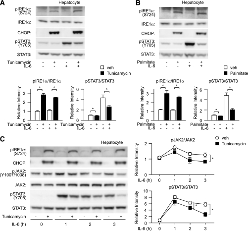
Induction of ER stress decreases tyrosine phosphorylation of hepatic STAT3.
- A and B, top: Wild-type mouse–derived hepatocytes were pretreated with tunicamycin or palmitate followed by stimulation with IL-6 and analyzed for expression of CHOP, phosphorylation of IRE1α, and tyrosine phosphorylation of STAT3.
Expansion of CD34+ cells in serum free medium
- Isolated CD34+ cells were seeded at a density of 5×10⁴cells per 500 µl of medium'>Stempro medium , in 24-well tissue culture plates .
- Growth factors used were IL-6, SCF, TPO, and Flt-3-L at a final concentration of 25 ng/ml with (test cells) and without (control cells) the addition of either zVADfmk −100 nM or zLLYfmk −10 µM (MP, , ).
- After the 10th day of culture, the cells were collected from the suspension culture, and centrifuged (1000 rpm for 5 minutes).
- The cells were used for assessing the
in vitro homing properties or were used forin vivo homing studies in NOD/SCID mice.
Functional assays
- For cytokine signalling, LN T cells (5 × 10⁶ cells/ml) were cultured 16 h with recombinant mouse IL-7 (10 ng/ml), IL-4 (40 ng/ml), IL-6 (45 ng/ml& ) or IL-15 (100 ng/ml& ) as previously described (
Retroviral transduction
-
Murine G0S2-V5 tagged cDNA was cloned into the MIGR1 retroviral vector
5′-CGAGCAATCAA GGAGCTAT-3′ ; mouse sh2,5′-GAGTCACATGCTGTTTCAA-3′ . - HEK 293T cells were cotransfected with a plasmid containing the retroviral vectorand ψ–ecotropic envelope.
- After 2 days of transfection, supernatant containing retrovirus was passed through a 0.4 µm filter before transduction.
- BM cells were flushed out from the femur and tibias of mice previously treated with 5-FU (150 mg/Kg) and cultured for two days in the presence of stem cell factor (100 ng/ml), IL-3 (6 ng/ml) and IL-6 (10 ng/ml) in X-vivo 15 medium .
- Cells were then transduced twice by spinoculation (60 min at 456 RCF) in the presence of polybrene (8 µg/ml).
- Next, 1×10⁶ BM cells were injected intravenously into the lateral tail vein of recipient mice that were previously irradiated with 950.
- NIH3T3 cells were transduced with retroviral supernatant in the presence of 8 µg/ml polybrene.
- EL4 cells were…


