Human Interleukin-3 Recombinant
Categories: HematopoietinsIL-3 familyRecombinant Human Cytokines$70.00 – $2,700.00
Description
Accession
P08700
Source
Optimized DNA sequence encoding Human Interleukin-3 mature chain was expressed in Escherichia Coli.
Molecular weight
Native human Interleukin-3 is generated by the proteolytic removal of the signal peptide and propeptide, the molecule has a calculated molecular mass of approximately 15 kDa. Recombinant IL-3 is a monomeric protein consisting of amino 133 acid residue subunits, and migrates as an approximately 15 kDa protein under non-reducing conditions and reducing conditions in SDS-PAGE.
Purity
>97%, as determined by SDS-PAGE and HPLC
Biological Activity
The ED(50) was determined by the dose-dependentproliferation of Human TF-1 cells was foundless than0.1 ng/ml.
Protein Sequence
MSRLPVLLLL QLLVRPGLQA PMTQTTPLKT SWVNCSNMID EIITHLKQPP LPLLDFNNLN GEDQDILMEN NLRRPNLEAF NRAVKSLQNA SAIESILKNL LPCLPLATAA PTRHPIHIKD GDWNEFRRKL TFYLKTLENA QAQQTTLSLA IF
Endotoxin
Endotoxin content was assayed using a LAL gel clot method. Endotoxin level was found to be less than 0.1 ng/µg(1EU/µg).
Presentation
Recombinant Interleukin-3 was lyophilized from a 0.2 μm filtered PBS solution pH7.0.
Reconstitution
A quick spin of the vial followed by reconstitution in distilled water to a concentration not less than 0.1 mg/mL. This solution can then be diluted into other buffers.
Storage
The lyophilized protein is stable for at least years from date of receipt at -20° C. Upon reconstitution, this cytokine can be stored in working aliquots at2° -8° C for one month, or at -20° C for six months, with a carrier protein without detectable loss of activity. Avoid repeated freeze/thaw cycles.
Usage
This cytokine product is for research purposes only.It may not be used for therapeutics or diagnostic purposes.
Interactor
Interactor
Molecular function
Molecular function
Methods
2.4. Generation of Red Blood Cells Ex Vivo
-
CD34+ cells were cultured in a three-step procedure in a medium'>medium'>serum-free medium'>medium as previously described [4 CD34+ cells/mL were first cultured in medium'>IMDM medium'>medium supplemented either with 1% BSA or 1% human AB plasma, 10
μ g/mL insulin , and 120μ g/mL iron-saturated human transferrin . - In the first step (days 0–8), 2 × 10⁴/mL C34+ cells were cultured in the presence of 10−6 M hydrocortisone , 100 ng/mL stem cell factor (, , sur , ), 5 ng/mL IL-3 , and 3 IU/mL erythropoietin (kindly provided by).
- On day 4, one volume of cell culture was diluted in four volumes of fresh medium containing hydrocortisone, SCF, IL-3, and erythropoietin.
- In the second step (days 8–10), the cells were replated at 5 × 10⁴/mL and cocultured on an adherent stromal layer in fresh medium supplemented with…
Cells
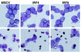
- Cell lines or primary cells were transduced with the indicated constructs using pMSCV-IRES-GFP-based retroviral vectors for 72 hr in the presence of IL-3, IL-6, SCF, and 7 μg/ml polybrene.
- For transient monobody expression, 1 × 10⁶ K562 cells were transfected with 5 μg of expression vectors encoding GFP fusion constructs of HA4-7c12 and mutants thereof using the Nucleofector Kit V , applying program T-016.
- Samples were harvested 48 hr later.
- Primary CML cells were isolated from bone marrow or peripheral blood samples of patients with Ph-chromosome-positive CML at time of diagnosis.
- All patients gave written informed consent before blood or bone marrow was obtained.
- Cells were transduced with concentrated lentiviral supernatants via spinoculation (1000 × g, 90 min) at a multiplicity of infection (moi) of 20 and cultivated in RPMI 1640 medium plus 10% FCS and 100 ng/ml human Interleukin 3 .
- The study was approved by the Institutional Review Board (IRB) of the Medical University of Vienna.
Processing whole blood samples
- Peripheral (PB.1 and PB.2) and cord (CB.1 and CB.2) blood-derived CD34+ cells were obtained from AllCells .
- Blood collections were performed at AllCells and using standard, 8 ml Vacutainer Cell Processing Tubes (both sodium citrate and sodium heparin-based tubes are acceptable ; , ).
- Appropriate documentation for informed consent was completed prior to blood collection .
- Vacutainers were processed within 24 hours of collection.
- Briefly, the PBMC-containing upper phase was collected and washed with ice-cold PBS .
- Cells were either frozen down or used directly for purification with the CD34 MicroBead Kit and used according to the manufacturer's protocol.
- Some samples were treated with Histopaque ( ; St. Louis, ) to minimize the number of red blood cells and centrifuged at 2000 rpm for 20 minutes without braking.
- The interface containing the PBMCs was removed if samples were treated with histopaque, cells washed again with chilled…
Erythroid precursor cultures
- After informed consent had been obtained, peripheral blood from CDA II patients and from 5 healthy control relatives was collected into sterile heparinised tubes.
- Light-density mononuclear cells obtained by centrifugation on density gradient were enriched for CD34+ cells by positive selection using CD34 microbeads according to the manufacturers' instructions.
- CD34+ cells were cultured at a density of 105 cells/mL in medium'>alpha-minimal medium'>essential medium (α-MEM; GIBCO) supplemented with 30% fetal bovine serum (FBS; GIBCO), as previously described [2 for 14 days; after 7 days of culture the medium was changed to ensure good cells feeding.
- Cell samples were collected on days 14 of culture (mature erythroblast stage) for further analysis.
Isolation and culture of cells
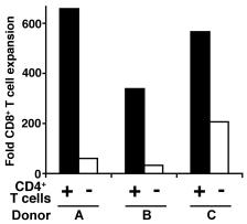
- Our procedures for isolation of subsets of Cs and T cells from blood have been described previously 6 cells/ml in complete medium (PMI 1640 Glutamax supplemented with 1% streptomycin and penicillin, 1% HEPES (all), 10% fetal bovine serum in the presence of recombinant human IL-3 (10 ng/ml& ) or GM-CSF (2 ng/ml, PeproTech).
- T cells were isolated from elutriated lymphocytes by negative selection and separation on.
- T cells were cultured at 10×106 cells/ml in complete medium and rested overnight before use.
CB collection and CD34+ cell isolation
- Umbilical CB samples from healthy newborns were obtained from the Andalusian Public Cord Blood Bank upon approval by our local (University of Granada) and Board Committee (ABR/JFJ/S-23).
- All human samples were obtained upon informed consent given by the parents.
- Mononuclear cells were isolated using Ficoll-Hypaque (GE Healthcare, Stockholm, Sweden).
- After lysing the red blood cells (Lysis solution , , ), CD34+ cells were purified by magnetic bead separation using the human CD34 MicroBead kit and the AutoMACS Pro separator as per manufacturer's instructions + cells were plated in liquid culture: Span medium ( Cell ) supplemented with SCF (100 ng/mL), FLT3L (100 ng/mL) and IL-3 (10 ng/mL)
Purification and Transduction of Mouse and Human Hematopoietic Progenitors
- Mouse Hematopoietic Progenitors (mLin−) were purified from BM mononuclear cells of C57BL6/6J mice byCell Depletion .
- The mouse cells were then prestimulated in StemSpam SFEM (serum-free expansion medium, StemCells Technologies, Canada) with 50ng/mL mouse Stem Cell Factor (mSCF) , 100ng/mL human Interleukin-11 (hIL-11), 100ng/mL human Flt-3 Ligand (hFlt-3L) and 10ng/mL human Interleukin-3 (hIL-3) (ocky , , ) for 4-6 hours.
- After which, mLin− cells were transduced with the different lentivirus.
- On the other hand, cord blood was collected from mothers attending the Royal Hospital, , after informed consent and via a protocol approved by the Local Research Ethics.
- Mononuclear cells (MNC) were obtained by Ficoll density centrifugation and ammonium chloride red cell lysis.
- Density-separated CB MNCs were depleted for lineage marker positive cells via theSep™ system ( Cell , ) according to the manufacturer’s instructions to generate Lineage negative (Lin−)…
Cell Culture
- The multiple myeloma cell line RPMI 8226, acute myeloid leukemia cell lines MOLM-13, and the erythroleukemia subtype of K562 were obtained from ATCC.
- KMS-12-PE-luc was generated in our laboratory by transduction of KMS-12-PE with a™ firefly luciferase control lentivirus obtained from™.
- Cord blood was obtained from Cleveland Cord Blood Center.
- Cells obtained from cord blood were kept in IMDM supplemented with 10% fetal bovine serum, penicillin G (50 units/ml), streptomycin (50 units/ml), and 10 ng/ml of the following cytokines, obtained from: Interleukin-3, interleukin-6, stem cell factor, and FLT3 ligand.
- All other cells were kept in RPMI 1640 (NaCl 103.45 mM, NaCO3 23.81 mM, Na2HPO4 5.63 mM, KCl 5.33 mM, Ca(NO3)2 4H2O 0.424 mM, MgSO4 0.407 mM, pH around 7.2), supplemented with 10% fetal bovine serum, penicillin G (50 units/ml), and streptomycin (50 units/ml).
- All cells were cultured at 37°C, 5% CO2, and humidified air.
- Cells were counted and viability assessed…
Culture of human mast cells
- Human umbilical cord blood was collected from mothers who had normal uncomplicated deliveries at Tufts Medical Center.
- Human cord blood-derived cultured mast cells (hCBMCs) were prepared using hematopoetic stem cells (C34+) isolated by positive selection of C34+/AC133+ cells by magnetic cell sorting using an AC133+ cell isolation kit as previously reported [+ cells were grown in medium'>medium'>medium'>serum-free expansion medium'>medium , supplemented with 100 ng/ml recombinant human stem cell factor (rhSCF; kindly supplied by Orphan , , ), 100 U/ml penicillin, 100 μg/ml streptomycin and IL-3 for the first 3 weeks, then in the medium'>medium'>medium'>serum-free expansion medium'>medium with 50 ng/ml IL-6 (ocky , , ) and for 8 to 10 weeks, with fetal bovine serum (/Gibco, , , ) added from week 6.
- The purity of the hCBMCs was…