Mouse Interleukin-10 Recombinant
Categories: Interferon-IL10 familyRecombinant Mouse Cytokines$70.00 – $4,700.00
Description
Accession
P18893
Source
Optimized DNA sequence encodingMouse Interleukin-10 mature chain was expressed in Escherichia Coli.
Molecular weight
Native MouseInterleukin-10 is generated by the proteolytic removal of the signal peptide and propeptide, the molecule has a calculated molecular mass of approximately kDa. Recombinant Mouse IL-10 is a monomer protein consisting of161 amino acid residue subunits, and migrates as an approximately19 kDa protein under non-reducing andreducing conditions in SDS-PAGE.
Purity
>95%, as determined by SDS-PAGE and HPLC
Biological Activity
ED50 was determined by thedose-dependent (with IL-4)proliferation of murine MC/9 cells, andwas found to be < ng/ml, corresponding to a specific activity of > x units/mg.
Protein Sequence
MPGSALLCCL LLLTGMRISR GQYSREDNNC THFPVGQSHM LLELRTAFSQ VKTFFQTKDQ LDNILLTDSL MQDFKGYLGC QALSEMIQFY LVEVMPQAEK HGPEIKEHLN SLGEKLKTLR MRLRRCHRFL KCENKSKAVE QVKSDFNKLE DQGVYKAMNE FDIFINCIEA YMMIKMKS
Endotoxin
Endotoxin content was assayed using a LAL gel clot method. Endotoxin level was found to be less than 0.1 ng/µg(1EU/µg).
Presentation
Recombinant Interleukin-10 was lyophilized from.2 μm filtered PBS solution, pH7.0.
Reconstitution
A quick spin of the vial followed by reconstitution in distilled water to a concentration not less than 0.1 mg/mL. This solution can then be diluted into other buffers.
Storage
The lyophilized protein is stable for at least years from date of receipt at -20° C. Upon reconstitution, this cytokine can be stored in working aliquots at2° -8° C for one month, or at -20° C for six months, with a carrier protein without detectable loss of activity. Avoid repeated freeze/thaw cycles.
Usage
This cytokine product is for research purposes only.It may not be used for therapeutics or diagnostic purposes.
Molecular function
Methods
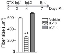
Forced premature expression of the antiinflammatory cytokine IL-10 impairs normal muscle repair.
- Recombinant IL-10 or IGF-1 (used as a control) was injected twice during the early stages of regeneration after CTX injury (Inj.).
- Induction of Alternatively Activated RAW Macrophages by Th2 Cytokines: The RAW264.7 cell line (1×10⁵) was treated with a combination of 10 ng/mL each of IL-4, IL-10, and IL-13 for 72 h, with the cytokine mixture added every 24 h. The resulting cells were macrophages in alternative activation state, named as IL-treated RAW macrophages.
Mice, BMDM preparation, and stimulation
- C57BL/6J wild type and C57BL/6J Cot/tpl2 KO animals were produced from heterozygous mice (Cot/tpl2 KO+/− × Cot/tpl2 KO+/−), and C57BL/6J Cot/tpl2 KD animals were produced from heterozygous C57BL/6J Cot/tpl2 KD+/− mice.
- All animals received care according to methods approved under institutional guidelines for the care and use of laboratory animals in research.
- BMDM were obtained as previously described (
- PD 0325901 and rapamycin were gifts from, respectively, Philip Cohen (Dundee, Scotland) and Victor Calvo .
Determination of cytokine activity
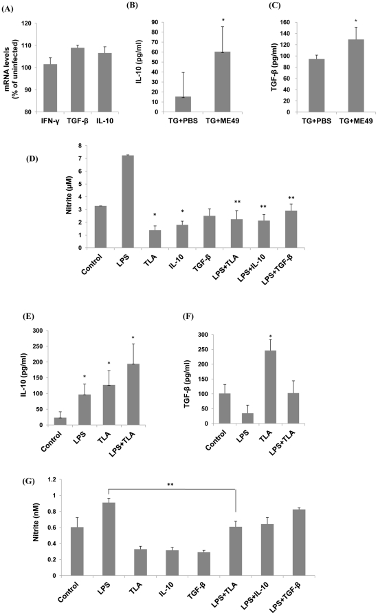
- Cytokine production in RAW 264.7 macrophages was examined using the following methodology: murine spleen cells were cultured in medium'>DMEM medium 0.5% FBS (starvation medium) overnight, followed by treatment with and without lipopolysaccharide and LCS101 for 24 hours.
- The plates were centrifuged and the supernatant analyzed for cytokine secretion.
- Cytokine levels were measured using murine IL-10 and TNF-α ELISA development kits and mouse IFN-γ using anElipair kit .
Macrophage polarization
- After 8 days, differentiated macrophages were harvested, counted and suspended in R10 + 15% LCM.
- Cells (3.5x10⁵ cells/well) were plated in 24-wells plates and kept overnight in order to adhere again.
- The next day, differentiated macrophages were polarized for 24h with interferon-γ (IFNγ) (100U/ml ) for M1, interleukin (IL)-4 (20ng/ml) for M2a or IL-10 (10ng/ml& ) for M2c macrophages.
- Non-polarized cells are referred to as M0 macrophages.
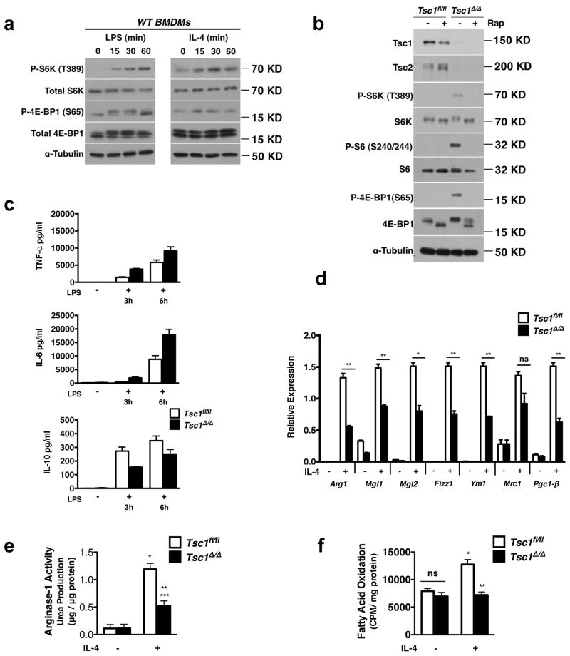
Tsc1 BMDMs Have Defective M2 Polarization and Enhanced Responses to LPS stimulation a. Immunoblot analysis of WT BMDMs stimulated with LPS or IL-4 for 15–60 min as indicated.
- c. Measurement of TNF-α, IL-6, and IL-10 secretion by ELISA after treatment with LPS for 3h and 6h, (n=2 representative experiments).
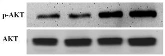
Concentration-dependent phosphorylation of AKT/STAT3 by IL-10 in HSFs.
- After a 30-min incubation with various doses of IL-10, cells were harvested and analyzed by Western blotting.
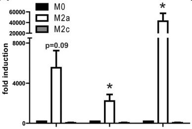
Induction of M2 macrophages.
- Gene expression analysis for M2 markers in M0 control, M2a and M2c bone marrow derived macrophages after 24 h of IL-4 (for M2a) or IL-10 (for M2c) polarization.