Description
Accession
O14788
Source
Optimized DNA sequence encoding Human RANK Ligand mature chain was expressed in Escherichia Coli.
Molecular weight
Native Human RANK Ligand is a type II transmembrane protein containing an extracellular TNF binding domain, the TNF binding domain has a calculated molecular mass of approximately 20 kDa. Recombinant RANKL is a monomeric protein consisting of 176 amino acid residue subunits. Recombinant RANKL migrates as an approximately 20 kDa protein under non-reducing conditions and reducing conditions in SDS-PAGE.
Purity
>98%, as determined by SDS-PAGE and HPLC
Biological Activity
The ED(50) determined by the dose-dependent stimulation of IL-8 production in Human PBMCs is ≤8 ng/ml.
Protein Sequence
MRRASRDYTK YLRGSEEMGG GPGAPHEGPL HAPPPPAPHQ PPAASRSMFV ALLGLGLGQV VCSVALFFYF RAQMDPNRIS EDGTHCIYRI LRLHENADFQ DTTLESQDTK LIPDSCRRIK QAFQGAVQKE LQHIVGSQHI RAEKAMVDGS WLDLAKRSKL EAQPFAHLTI NATDIPSGSH KVSLSSWYHD RGWAKISNMT FSNGKLIVNQ DGFYYLYANI CFRHHETSGD LATEYLQLMV YVTKTSIKIP SSHTLMKGGS TKYWSGNSEF HFYSINVGGF FKLRSGEEIS IEVSNPSLLD PDQDATYFGA FKVRDID
Endotoxin
Endotoxin content was assayed using a LAL gel clot method. Endotoxin level was found to be less than 0.1 ng/µg(1EU/µg).
Presentation
Recombinant RANKL was lyophilized from a 0.2 μm filtered PBS solution pH7.5.
Reconstitution
A quick spin of the vial followed by reconstitution in distilled water to a concentration not less than 0.1 mg/mL. This solution can then be diluted into other buffers.
Storage
The lyophilized protein is stable for at least years from date of receipt at -20° C. Upon reconstitution, this cytokine can be stored in working aliquots at2° -8° C for one month, or at -20° C for six months, with a carrier protein without detectable loss of activity. Avoid repeated freeze/thaw cycles.
Usage
This cytokine product is for research purposes only.It may not be used for therapeutics or diagnostic purposes.
Biological Process
Molecular function
Molecular function
Molecular function
Methods
In vitro differentiation of human osteoclasts and confocal microscope analysis
- Human osteoclasts were generated by culture of peripheral blood monocytes with M-CSF and RANKL using a standard protocol.
- Peripheral blood mononuclear cells (PBMCs) were isolated from heparinized blood samples by Ficoll density gradient centrifugation .
- PBMCs were cultured either on glass coverslips for differentiation analysis or on dentine discs for resorption assays in alpha MEM , 10% FCS , 1% Ultraglutamine , 1% Pen/Strep, 25 ng/mL human M-CSF , and 30 ng/mlankl (ocky , , ).
- Cells were cultured for 2 weeks with medium changes twice weekly.
- Cells on coverslips were fixed in 4% paraformaldehyde (PFA) in 1× phosphate-buffered saline (PBS) and then stained with Phalloidin-Alexa 488 , DAPI , and tartrate-resistant acid phosphatase (TRAP) activity using Naphtol-AS-MX-Phosphate and Fast-Red-Violet LB .
- TRAP-positive cells with ≥3 nuclei and actin rings were counted as osteoclasts.
- Dentine discs were cleaned with 1% SDS and resorption pits were visualized by black…
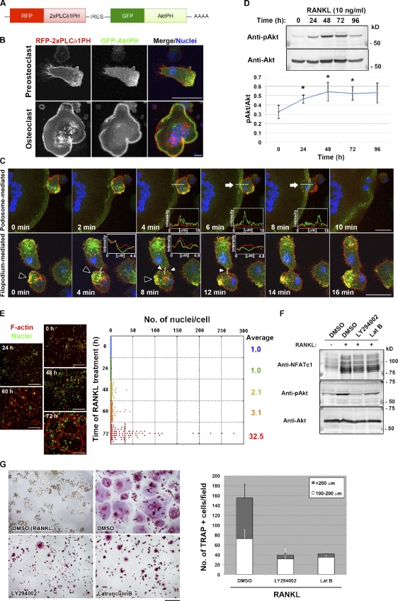
Polarized membrane extensions mediate osteoclast fusion.
- (D, top) Immunoblot analysis of lysates of RAW264.7 macrophages stimulated with RANKL for the indicated times with antibodies to Ser473-phosphorylated (p) or total forms of Akt.
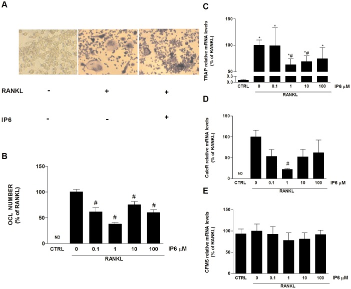
Inositol hexakisphosphate (IP6) directly inhibits osteoclast formation induced by RANKL.
- RAW 264.7 cells cultured for 5 days with no stimulation with RANKL (left).
Cell isolation and culture
- The primary human material was obtained after written informed consent and with approval of the Medical Ethical Committee of the Academic Medical Centre (Amsterdam, The Netherlands), which specifically approved this study.
- Mobilized peripheral blood samples were harvested from leukapheresis material of G-CSF-treated mantle cell lymphoma patient treated with chemotherapy and G-CSF (2×5 µg/kg/day subcutaneously, , , ).
- These patients are in remission and therefore the CD34+ cells are probably normal.
- The samples were diluted in phosphate-buffered saline (PBS; Fresenius Kobi's, The ) with 2 mM ethylenediamine tetra acetic acid (EDTA) and 0.5% w/v bovine serum albumin (BSA, , ).
- CD34+ cells were obtained by magnetic cell sorting (MACS) reaching purities of 95% or more.
- Neutrophils were isolated from the heparinized blood of healthy human subjects with a Ficoll-Paque PLUS (GE Healthcare, Uppsala, Sweden) gradient and subsequent lysis of erythrocytes as described 2, 10 µg/ml) and harvested at day…
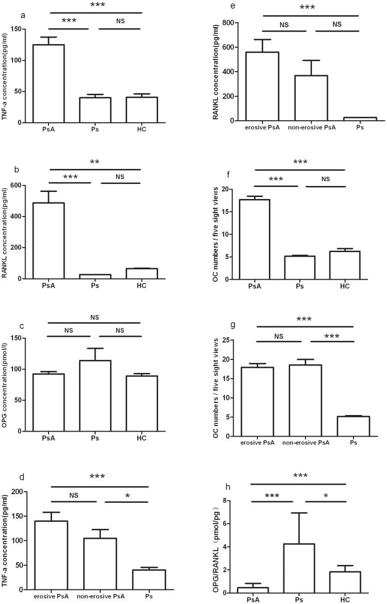
Soluble mediators of bone remodeling in the circulation of patients with PsA.
- Bar plots show mean concentrations with SEM (standard error of the mean) of (a) TNF-α, (b) RANKL, (c) OPG, (f) OCs and (h) OPG/RANKL (pmol/l to pg/ml) in healthy controls (HCs), patients with psoriasis (Ps) and patients with PsA.
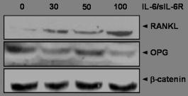
The effect of the IL-6/sIL-6R complex on RANKL, JAK2, STAT3 in fibroblast-like synoviocytes (FLS).
- Stimulation of FLS with IL-6 and sIL-6R at several different concentrations (0, 30, 50, 100 ng of both) for 30 minutes induced RANKL protein expression in a dose-dependent manner.
Osteoclastogenesis assays
- Freshly isolated, peripheral blood monocytes were obtained from human donors, as previously described [5 cells per well in a 12-well plate containing 5×10⁶ preplated GCTB stromal cells, and monocytes were also plated alone (6.25×10⁵cells/well) in the presence of 25 ng/mL macrophage colony stimulating factor (M-CSF) with or without 40 ng/mL RANKL (both, , ) as negative and positive controls, respectively.
- In all cultures, media and cytokines were changed every 3-4 days.
- After 10 days of culture, cells were stained for tartrate resistant acid phosphatase (TRAP) using a commercially available kit and triplicate counts of TRAP-positive multinucleated giant cells were performed.
- Comparisons were analyzed statistically using the Mann–Whitney U test with post-hoc adjustment of the p value.
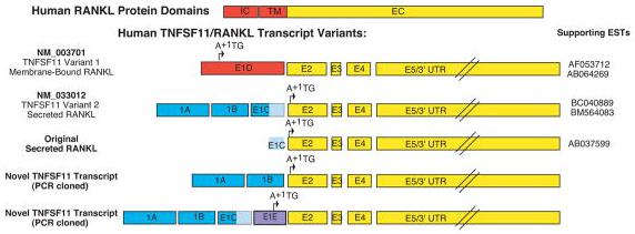
TNFSF11: mRNA transcript variants, genomic loci and sequence conservation A.
- Color coded schematics showing the exon structure for the human TNFSF11 mRNA transcripts aligned with the known RANKL protein domains.
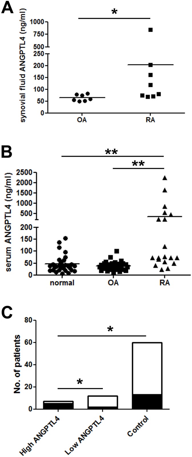
Serum and synovial fluid ANGPTL4 concentrations are elevated in RA.
- Serum from ‘high ANGPTL4’ RA patients is more likely to have detectable RANKL (black shading; RANKL-positive) than either serum from ‘low ANGPTL4’ RA patients or controls.