
Mouse SDF-1 alpha Recombinant
Categories: CXC chemokinesRecombinant Mouse Cytokines$70.00 – $2,700.00
Description
Accession
P40224
Source
Optimized DNA sequence encoding Mouse SDF-1 alpha mature chain was expressed in Escherichia Coli.
Molecular weight
Native Mouse Stromal Cell-Derived Factor-1 alpha, generated by the proteolytic removal of the signal peptide and propeptide,the molecule has a calculated molecular mass of approximately 8 kDa. Recombinant SDF-1 alpha is a monomer protein consisting of 69 amino acid residue subunits, migrates as an approximately 8 kDa protein under non-reducing and reducing conditions in SDS-PAGE.
Purity
>95%, as determined by SDS-PAGE and HPLC
Biological Activity
Determined by its ability to chemoattract human peripheral T cells activated with PHA and IL-2 using a concentration range of-40 ng/ml.
Protein Sequence
MDAKVVAVLA LVLAALCISD GKPVSLSYRC PCRFFESHIA RANVKHLKIL NTPNCALQIV ARLKNNNRQV CIDPKLKWIQ EYLEKALNK
Endotoxin
Endotoxin content was assayed using a LAL gel clot method. Endotoxin level was found to be less than 0.1 ng/µg(1EU/µg).
Presentation
Recombinant SDF-1 alpha was lyophilized from a 0.2 μm filtered solution in.5% glycine,.5% sucrose,.01% Tween80, mM Glutamic acid, pH.5.
Reconstitution
A quick spin of the vial followed by reconstitution in distilled water to a concentration not less than 0.1 mg/mL. This solution can then be diluted into other buffers.
Storage
The lyophilized protein is stable for at least years from date of receipt at -20° C. Upon reconstitution, this cytokine can be stored in working aliquots at2° -8° C for one month, or at -20° C for six months, with a carrier protein without detectable loss of activity. Avoid repeated freeze/thaw cycles.
Usage
This cytokine product is for research purposes only.It may not be used for therapeutics or diagnostic purposes.
Interactor
Q8JLJ0
Interactor
Q8QMN0
Interactor
Q9JFT2
Biological Process
Molecular function
Molecular function
Methods
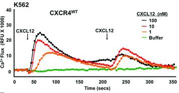
WHIM mutation CXCR4R334X increases CXCL12-induced calcium flux response, but allows homologous desensitization: analysis of transfected cells.
- The cell line, receptor and CXCL12 doses are indicated at the top left and right of each panel.
ELISA
- The concentrations of TNFα, CXCL12 and CCL2 were measured in cell supernatants by using ELISA kits in accordance with the manufacturer's protocols (TNFα and CXCL12 from R&D systems; CCL2 from) on an ETIMax-3000 reader .
- Supernatants were collected 24 h after infection, spun at 10,000 rpm for 10 min to discard bacteria and cell debris, and stored at −80°C until assayed.
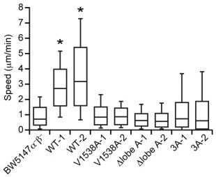
The expression of the Δlobe A and 3A mutant fails to restore motility and polarity in BW5147α–β– cells.
- BW5147α–β– cells stably expressing WT or mutant DOCK2 (V1538A, Δlobe A, or 3A) were stimulated in suspension with CXCL12, and stained with phalloidin and DAPI.
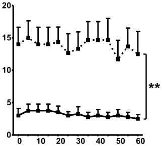
Dasatinib inhibits actin polymerisation and migration of CLL cells in response to CXCL12.
- A: Analysis of the effect of dasatinib on actin polymerization following CXCL12 treatment of CLL cells.
Cell Migration
- The upper chamber wells of CIM-Plates 16 were precoated with fibronectin, added to wells at 10 µg/ml (30 µl) and removed after 30 min.
- The lower chamber wells were filled with 160 µl culture medium, allowing a clearly-defined meniscus to form.
- SDF1, MCP1 and TNFα were added to the lower chambers at a final concentration of 100 ng/ml.
- CIM-Plates were equilibrated for at least 30 min in a culture hood at room temperature.
- Trypsin-EDTA-dispersed MPCs (100 µl in complete medium) were then plated in the fibronectin-precoated upper chamber wells at 306×103 cells/cm2 (600000 cells/ml) in 100 µl complete medium, which we determined as the optimal density to ensure confluent monolayers.
- The CIM-Plates were inserted into the cradle pockets of the RTCA DP Analyzer, and the background impedance of each well was measured after 1 h. The RTCA DP Analyzer, placed in a standard cell-culture incubator, monitored the migrated cell number…
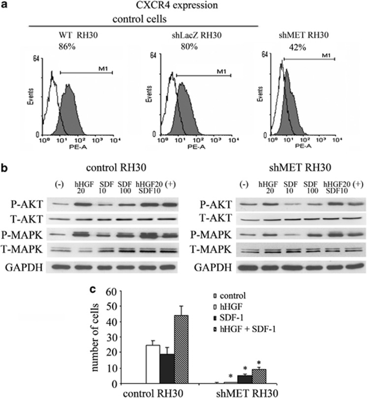
Expression and function of the CXCR4 receptor in MET-depleted cell.
- (b) Western blot analysis of AKT and MAPK activation in shMET RH30 cells showed a decrease in the phosphorylation of these kinases following stimulation with human HGF (20 ng/ml) and SDF-1 (10 ng/ml and 100 ng/ml).
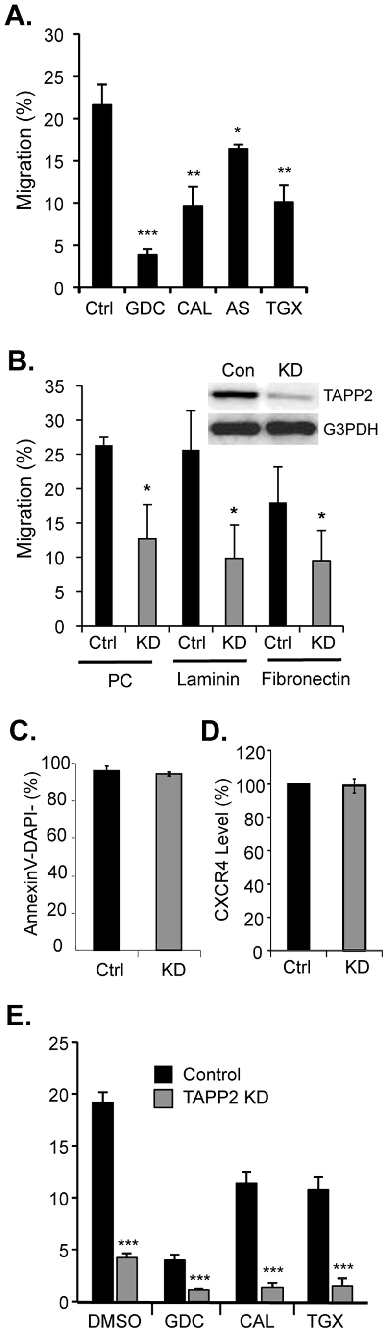
TAPP2 knockdown impairs SDF-1-induced migration.
- A NALM-6 cells migration towards SDF-1 is impaired by PI3K inhibitors.
Topical treatment of the small intestine with cytokines increases HPC-7 adhesion in vivo.
- No increase in adhesion was observed following topical treatment of the gut with CXCL12.
Topical treatment of the small intestine with cytokines increases HPC-7 adhesion in vivo.
- No increase in adhesion was observed following topical treatment of the gut with CXCL12.
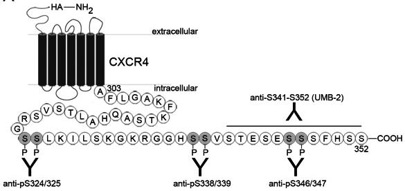
Specificity of phospho-sensitive anti-CXCR4 antibodies.
- B–G, HEK293 cells stably transfected with hemagglutinin epitope-tagged CXCR4 received CXCL12 (20 nM) or vehicle for 15 min and were lysed.